Most everyone who has had dental work, has experienced x-rays. Typically these have been done with the big swinging arm and an electronic sensor gently inserted into one’s mouth. The digital image which is captured can be stored on a computer, along with your dental records.
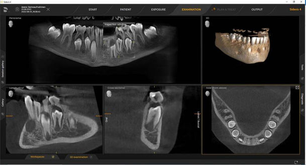
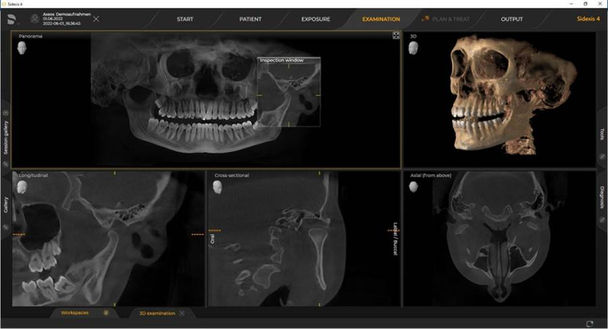
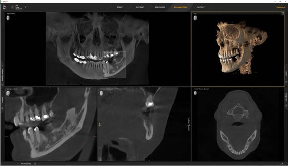
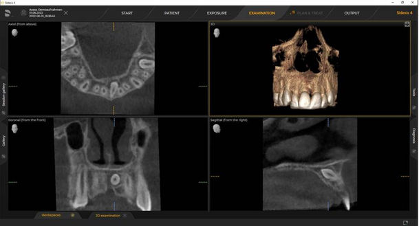
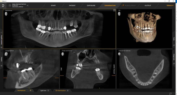
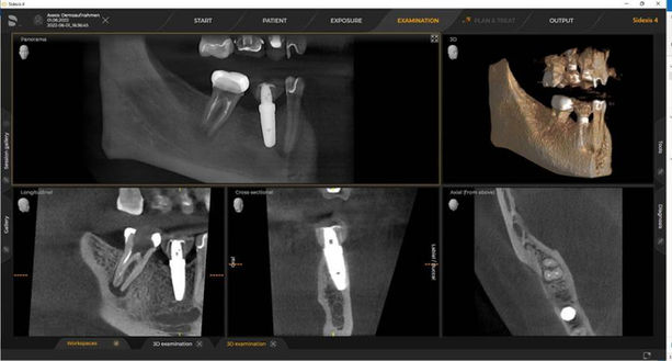
At Carson and Acasio we have taken digital technology to another level with the recent addition of the Dentsply Sirona Axeos CT scan system which can provide us with much more critical information for difficult cases.
X-rays, and CT scans, are essential diagnostic tools which provide valuable information for the prevention and restoration of dental issues. Our dentists and hygienists use this information to more accurately and effectively detect hidden dental abnormalities in order to frame your treatment plan.

Digital X-rays
& CT Scans
Let’s start with the basics. What are dental x-rays?
A dental x-ray is a digital image of the teeth, bones, soft tissue and gums which help us find problems such as cavities, teeth which have not erupted through the gums, and bone loss which we can’t see through a visual examination. We may also use x-rays following a procedure to confirm the remedial work has been performed correctly.
For most evaluations, we offer four types of dental x-rays:

Bitewings.
These show the upper and lower back teeth in the same image. They can expose any decay and how the teeth align when you bite down.

Periapical.
These x-rays show an entire tooth from the top to the root and bone which supports the tooth. We look for issues below the gumline which we can’t see … like cysts, bone loss and impacted teeth.

Occlusal.
These show the top or bottom of the mouth where we can find potential extra teeth, or teeth which have not yet broken through the gum line. We can also discover jaw fractures, cleft palate issues, and other growths.

Panoramic.
These x-rays allow us to see the entire lower jaw. We can see any number of problems from bone abnormalities to cysts, growths, and bite and jaw issues.
By contrast, a dental CT scan provides a more detailed image of dental structures, soft tissues, nerve pathways and bone in just one scan.

We can take a 360-degree image of one’s head which can help us to diagnose diseases of the jaw, bony structures of the face, nasal cavity and sinuses. We use a CT scan to locate impacted teeth, diagnose TMJ (temporomandibular joint) disorder, ensure accurate placement of dental implants, evaluate bone structure and more.
Our CT scan system works with much the same low radiation level technology as used by physicians worldwide to diagnose and treat major health issues.

